Vasculitis involves inflammation of blood vessels. This inflammation targets the vessel walls directly. As swelling increases, vessel integrity weakens. Blood flow becomes irregular or restricted. Some vessels may narrow or completely close. Others balloon outward or rupture. The effects depend on vessel size and location. Any organ can be involved. Symptoms vary accordingly. Without circulation, tissue begins to suffer. Organ damage may follow. Early recognition is essential for intervention.
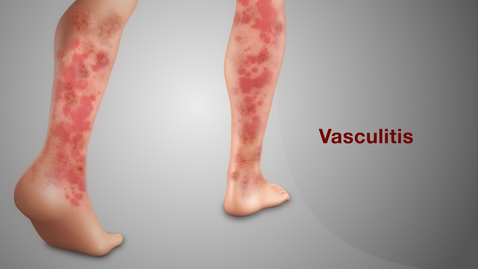
Small-vessel involvement often causes skin rashes, nerve tingling, or kidney dysfunction
Small vessels supply skin, nerves, and kidneys. Their inflammation produces specific symptoms. Skin may develop purpura or red spots. These lesions don’t fade when pressed. Nerves become irritated or deprived of oxygen. Tingling, numbness, or weakness may result. In the kidneys, vessels filter blood poorly. Waste products remain elevated. Urine changes color or volume. These signs often appear first. Blood and urine tests support diagnosis. Biopsy confirms tissue changes under microscopy.
Medium-vessel vasculitis may affect the gut, muscles, or peripheral nerves unexpectedly
Medium vessels reach deeper tissues. Gut involvement brings pain after meals. Blood flow fails to meet demand. Cramping becomes severe. Some patients report nausea or weight loss. Muscles may feel weak or tender. Peripheral nerves lose function. Motor control decreases in affected areas. These changes confuse diagnosis. Initial symptoms mimic more common disorders. Only detailed investigation clarifies the pattern. Medium-vessel disease progresses quickly. Prompt treatment reduces permanent damage risks.
Large-vessel inflammation can impair major arteries supplying limbs or critical organs
Large-vessel vasculitis affects aorta and branches. These include arteries to limbs and brain. Inflammation narrows their diameter. Reduced blood flow limits function. Arms tire easily. Legs may ache when walking. Some patients feel lightheaded or dizzy. Vision may blur during flares. When arteries thicken, pulses weaken. Bruits or vascular sounds are heard during exams. Imaging reveals wall thickening or stenosis. CT angiography confirms extent of disease. Intervention depends on affected arteries.
Systemic symptoms like fatigue and fever often precede organ-specific complications
Many cases begin subtly. Fatigue appears without explanation. Fever may rise intermittently. Appetite fades. Weight loss occurs over weeks. These signs resemble infection or autoimmunity. No single symptom confirms vasculitis. Combined patterns raise suspicion. Laboratory markers of inflammation increase. CRP and ESR often rise. Autoantibodies may be detected. Still, results vary widely. Physicians consider clinical context. Symptoms evolve in clusters. Careful history reveals overlooked signs. Patterns eventually guide diagnosis direction.
Diagnosis involves imaging, lab markers, and tissue biopsy for histological confirmation
Blood tests suggest inflammation. But they rarely confirm vasculitis alone. Imaging becomes crucial. PET scans highlight vessel inflammation. CT angiography shows vessel changes. MRI provides tissue detail. Biopsy remains gold standard. Sampled tissue reveals damaged vessels. Inflammatory cells cluster near walls. Necrosis may appear in severe cases. Biopsy location depends on symptoms. Skin, kidney, or temporal artery are common targets. Diagnostic delay worsens outcomes. Multimodal approach improves accuracy.
Treatment suppresses immune activity to prevent further vessel damage and organ failure
The immune system drives vasculitis. Treatment focuses on suppression. Corticosteroids start quickly. They reduce inflammation rapidly. High doses are often needed initially. Immunosuppressants follow to maintain control. Methotrexate, azathioprine, or cyclophosphamide may be used. Biologics like rituximab offer targeted therapy. Duration of treatment varies. Relapses are common. Long-term monitoring is essential. Dosage adjustments respond to disease activity. Early withdrawal increases risk of flare. Consistent follow-up ensures disease stability.
Chronic vasculitis may lead to scarring, aneurysm formation, or permanent tissue loss
Uncontrolled vasculitis causes damage. Vessel walls may scar. Elasticity disappears. Blood vessels become brittle. Aneurysms can form in weakened areas. Rupture risk increases. Some organs lose tissue permanently. Kidney scarring reduces filtration. Lung tissue may become fibrotic. Neurological function doesn’t always return. Prevention of these outcomes drives aggressive care. Stabilization, not reversal, becomes the goal. Early treatment avoids irreversible injury. Chronic cases need customized support plans.
Specific types like Takayasu, GPA, or Behçet’s follow different vascular and organ patterns
Vasculitis includes many types. Each targets different vessels. Takayasu affects large arteries. Usually young women are diagnosed. Granulomatosis with polyangiitis affects lungs, kidneys, and sinuses. Behçet’s includes ulcers and vascular clots. Diagnosis requires understanding patterns. Geographic and ethnic distribution varies. Genetic factors contribute. Disease behavior changes over time. Even within a type, symptoms differ. Treatment protocols are similar but personalized. Classification supports clearer expectations and research.
Long-term outcomes depend on organ damage, treatment adherence, and flare prevention
Some patients enter remission. Others face recurring flares. Outcomes vary widely. Early control improves outlook. Organ preservation is key. Monitoring tracks complications. Blood pressure, kidney function, and oxygenation must remain stable. Lifestyle affects recovery. Smoking worsens vascular health. Exercise enhances circulation. Support groups offer stability. Mental health requires attention. Chronic disease alters life structure. Success comes with patience and informed care. Each flare teaches new lessons.